Best Disease - 61 Year Old Female
|
|
61-year-old woman was seen in the office on 4/11/11. She was diagnosed with Best’s disease about 30 years ago. She and her many family members were tested, both at the VA Hospital and then at USF. Since then her vision is reasonable. She does note it gradually getting a little bit worse, especially the last two years. She has been diabetic for four years. She has a history of high blood pressure.
VISUAL ACUITY: OD 20/50, OS 20/50. IOP: OD 12, OS 14. There is 1+ nuclear sclerosis in both eyes.
EXTENDED OPHTHALMOSCOPY:
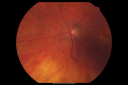
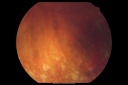
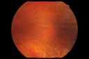
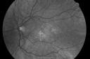
OD: Vertical C/D ratio is 0.5. There is posterior vitreous separation. There are yellow pigment spots around the fovea. There are also punched-out areas of chorioretinal atrophy in the periphery.
OS: Vertical C/D ratio is 0.5. There is posterior vitreous separation. There are yellow pigment spots around the fovea and chorioretinal atrophy in the periphery.
OCT SCAN: The OCT scan shows no intraretinal or subretinal fluid in either eye. There is a loss of normal retinal architecture.
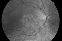
Photos confirm clinical findings.
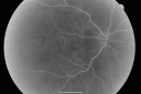
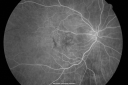
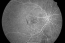
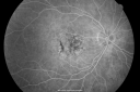
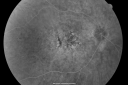
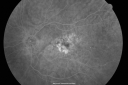
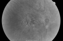
FLUORESCEIN ANGIOGRAM: Shows areas of hyperfluorescence in the right eye early on, corresponding to mild nongeographic atrophy. There is also hypofluorescence corresponding to pigment clumping. The left eye has geographic atrophy just inferonasal to the fovea, which touches the fovea.
IMPRESSION:
1. AGE-RELATED MACULAR DEGENERATION – BOTH EYES
2. HISTORY OF BEST’S DISEASE
3. PERIPHERAL CHORIORETINAL ATROPHY – BOTH EYES
DISCUSSION: I explained to the patient she does have macular disease in both eyes, which is consistent with Best’s disease, although at this point it does look like macular degeneration. I asked her to take eye vitamins, which she is doing, to follow her vision, follow the grid, and return urgently should she notice a change in either eye. She will also return here for a check in about one year.
|

Best Disease for 30 Years - 20/50 Vision both eyes - 61 Year Old Womanvista 902 veces61-year-old woman was diagnosed with Best’s disease about 30 years ago. She and her many family members were tested, both at the VA Hospital and then at USF. OD 20/50, OS 20/50
(0 votos)
|
|
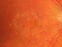
Best Disease for 30 Years - 20/50 Vision both eyes - 61 Year Old Womanvista 771 veces61-year-old woman was diagnosed with Best’s disease about 30 years ago. She and her many family members were tested, both at the VA Hospital and then at USF. OD 20/50, OS 20/50
(0 votos)
|
|

Best Disease for 30 Years - 20/50 Vision both eyes - 61 Year Old Womanvista 745 veces61-year-old woman was diagnosed with Best’s disease about 30 years ago. She and her many family members were tested, both at the VA Hospital and then at USF. OD 20/50, OS 20/50
(0 votos)
|
|
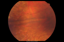
Best Disease for 30 Years - 20/50 Vision both eyes - 61 Year Old Womanvista 694 veces61-year-old woman was diagnosed with Best’s disease about 30 years ago. She and her many family members were tested, both at the VA Hospital and then at USF. OD 20/50, OS 20/50
(0 votos)
|
|
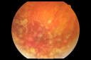
Best Disease for 30 Years - 20/50 Vision both eyes - 61 Year Old Womanvista 893 veces61-year-old woman was diagnosed with Best’s disease about 30 years ago. She and her many family members were tested, both at the VA Hospital and then at USF. OD 20/50, OS 20/50
(0 votos)
|
|

Best Disease for 30 Years - 20/50 Vision both eyes - 61 Year Old Womanvista 742 veces61-year-old woman was diagnosed with Best’s disease about 30 years ago. She and her many family members were tested, both at the VA Hospital and then at USF. OD 20/50, OS 20/50
(0 votos)
|
|
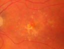
Best Disease for 30 Years - 20/50 Vision both eyes - 61 Year Old Womanvista 721 veces61-year-old woman was diagnosed with Best’s disease about 30 years ago. She and her many family members were tested, both at the VA Hospital and then at USF. OD 20/50, OS 20/50
(0 votos)
|
|
Best Disease for 30 Years - 20/50 Vision both eyes - 61 Year Old Womanvista 640 veces61-year-old woman was diagnosed with Best’s disease about 30 years ago. She and her many family members were tested, both at the VA Hospital and then at USF. OD 20/50, OS 20/50
(0 votos)
|
|
Best Disease for 30 Years - 20/50 Vision both eyes - 61 Year Old Womanvista 670 veces61-year-old woman was diagnosed with Best’s disease about 30 years ago. She and her many family members were tested, both at the VA Hospital and then at USF. OD 20/50, OS 20/50
(0 votos)
|
|
Best Disease for 30 Years - 20/50 Vision both eyes - 61 Year Old Womanvista 668 veces61-year-old woman was diagnosed with Best’s disease about 30 years ago. She and her many family members were tested, both at the VA Hospital and then at USF. OD 20/50, OS 20/50
(0 votos)
|
|
Best Disease for 30 Years - 20/50 Vision both eyes - 61 Year Old Womanvista 640 veces61-year-old woman was diagnosed with Best’s disease about 30 years ago. She and her many family members were tested, both at the VA Hospital and then at USF. OD 20/50, OS 20/50
(0 votos)
|
|
Best Disease for 30 Years - 20/50 Vision both eyes - 61 Year Old Womanvista 617 veces61-year-old woman was diagnosed with Best’s disease about 30 years ago. She and her many family members were tested, both at the VA Hospital and then at USF. OD 20/50, OS 20/50
(0 votos)
|
|
Best Disease for 30 Years - 20/50 Vision both eyes - 61 Year Old Womanvista 614 veces61-year-old woman was diagnosed with Best’s disease about 30 years ago. She and her many family members were tested, both at the VA Hospital and then at USF. OD 20/50, OS 20/50
(0 votos)
|
|
Best Disease for 30 Years - 20/50 Vision both eyes - 61 Year Old Womanvista 567 veces61-year-old woman was diagnosed with Best’s disease about 30 years ago. She and her many family members were tested, both at the VA Hospital and then at USF. OD 20/50, OS 20/50
(0 votos)
|
|
Best Disease for 30 Years - 20/50 Vision both eyes - 61 Year Old Womanvista 601 veces61-year-old woman was diagnosed with Best’s disease about 30 years ago. She and her many family members were tested, both at the VA Hospital and then at USF. OD 20/50, OS 20/50
(0 votos)
|
|
Best Disease for 30 Years - 20/50 Vision both eyes - 61 Year Old Womanvista 579 veces61-year-old woman was diagnosed with Best’s disease about 30 years ago. She and her many family members were tested, both at the VA Hospital and then at USF. OD 20/50, OS 20/50
(0 votos)
|
|
Best Disease for 30 Years - 20/50 Vision both eyes - 61 Year Old Womanvista 590 veces61-year-old woman was diagnosed with Best’s disease about 30 years ago. She and her many family members were tested, both at the VA Hospital and then at USF. OD 20/50, OS 20/50
(0 votos)
|
|
Best Disease for 30 Years - 20/50 Vision both eyes - 61 Year Old Womanvista 585 veces61-year-old woman was diagnosed with Best’s disease about 30 years ago. She and her many family members were tested, both at the VA Hospital and then at USF. OD 20/50, OS 20/50
(0 votos)
|
|
Best Disease for 30 Years - 20/50 Vision both eyes - 61 Year Old Womanvista 640 veces61-year-old woman was diagnosed with Best’s disease about 30 years ago. She and her many family members were tested, both at the VA Hospital and then at USF. OD 20/50, OS 20/50
(0 votos)
|
|
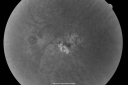
Best Disease for 30 Years - 20/50 Vision both eyes - 61 Year Old Womanvista 654 veces61-year-old woman was diagnosed with Best’s disease about 30 years ago. She and her many family members were tested, both at the VA Hospital and then at USF. OD 20/50, OS 20/50
(0 votos)
|
|
| 20 archivos en 1 página(s) |
|
61-year-old woman was seen in the office on 4/11/11. She was diagnosed with Best’s disease about 30 years ago. She and her many family members were tested, both at the VA Hospital and then at USF. Since then her vision is reasonable. She does note it gradually getting a little bit worse, especially the last two years. She has been diabetic for four years. She has a history of high blood pressure.
VISUAL ACUITY: OD 20/50, OS 20/50. IOP: OD 12, OS 14. There is 1+ nuclear sclerosis in both eyes.
EXTENDED OPHTHALMOSCOPY:
OD: Vertical C/D ratio is 0.5. There is posterior vitreous separation. There are yellow pigment spots around the fovea. There are also punched-out areas of chorioretinal atrophy in the periphery.
OS: Vertical C/D ratio is 0.5. There is posterior vitreous separation. There are yellow pigment spots around the fovea and chorioretinal atrophy in the periphery.
OCT SCAN: The OCT scan shows no intraretinal or subretinal fluid in either eye. There is a loss of normal retinal architecture.
Photos confirm clinical findings.
FLUORESCEIN ANGIOGRAM: Shows areas of hyperfluorescence in the right eye early on, corresponding to mild nongeographic atrophy. There is also hypofluorescence corresponding to pigment clumping. The left eye has geographic atrophy just inferonasal to the fovea, which touches the fovea.
IMPRESSION:
1. AGE-RELATED MACULAR DEGENERATION – BOTH EYES
2. HISTORY OF BEST’S DISEASE
3. PERIPHERAL CHORIORETINAL ATROPHY – BOTH EYES
DISCUSSION: I explained to the patient she does have macular disease in both eyes, which is consistent with Best’s disease, although at this point it does look like macular degeneration. I asked her to take eye vitamins, which she is doing, to follow her vision, follow the grid, and return urgently should she notice a change in either eye. She will also return here for a check in about one year.


