Fundus Photos - Angiogram - OCT - 69 Year Old Woman
|
|
69-year-old woman who I saw in the office on July 15, 2009. She had cataract surgery done in 2006, first in the left eye and subsequently in the right eye. She had vitreous separations with floaters. She also sees temporal flashing lights.
In addition she has an unusual symptom of seeing lights in both eyes, sort of blotchy lights that move around throughout the visual field that are there most of the time. They are not regionalized and they seem to be in both eyes and they are not associated with any sort of visual phenomenon, such as looking at street lights or headlights and they can happen in dim lighting or regular lighting.
VISUAL ACUITY: OD 20/50. Pinhole 20/30. OS 20/30. IOP: OD 16, OS 15. Amsler grid is normal in both eyes.
SLIT EXAMINATION: The right eye has a posterior chamber intraocular lens is in good position with clear capsular. The left eye has a posterior chamber intraocular lens is in good position with 2+ posterior capsular opacity and an open capsule center.
EXTENDED OPHTHALMOSCOPY:
OD: Vertical C/D ratio is 0.5. There is a posterior vitreous separation and trace epiretinal membrane.
OS: Vertical C/D ratio is 0.5. There is posterior vitreous separation and trace epiretinal membrane.
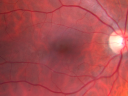
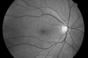
OCT SCAN: The OCT scan were normal as are the photos and fluorescein angiograms.
ATYPICAL PHOTOPSIAS
DISCUSSION: I explained to the patient that the photopsias she is seeing are atypical and do sound like something that we see occasionally in people who have retinal inflammatory disease, such as multiple evanescent white-dot syndrome or acute zonal occult outer retinopathy. All of those diseases though tend to have transient lights and they also tend to be regionalized. Her case is different in that she has had the lights now for several years and they seem to be diffuse.
There are some drugs which will cause retinal toxicity, such as Digitalis that can cause people to see unusual light phenomenon and that tends to be a passing thing and she is not on any medications that to my knowledge cause any retinal toxicities.
I suggested, just to be on the safe side she have a neurological evaluation, because occasionally visual symptoms are not from the eye, but from the visual pathways and it is possible that she has chronic irritation of her visual pathways somewhere.
If you have not done so, it would be reasonable to obtain a visual field, just to make sure there is not something going on with her visual pathways that might be knocking on her visual field and I told her she does not need to return here unless you or she notes further problems. Thank you for allowing me to participate in her care.
|
|
|
|
|
|
|
|
|
|
|
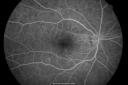
69 Year Old Woman Normal Fluorescein Angiogram967 views69-year-old woman OD 20/50 - OS 20/30 ATYPICAL PHOTOPSIAS

(0 votes)
|
|
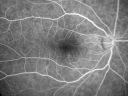
69 Year Old Woman Normal Fluorescein Angiogram858 views69-year-old woman OD 20/50 - OS 20/30 ATYPICAL PHOTOPSIAS
(0 votes)
|
|
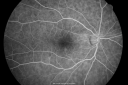
69 Year Old Woman Normal Fluorescein Angiogram901 views69-year-old woman OD 20/50 - OS 20/30 ATYPICAL PHOTOPSIAS
(0 votes)
|
|
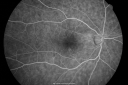
69 Year Old Woman Normal Fluorescein Angiogram751 views69-year-old woman OD 20/50 - OS 20/30 ATYPICAL PHOTOPSIAS
(0 votes)
|
|
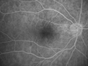
69 Year Old Woman Normal Fluorescein Angiogram759 views69-year-old woman OD 20/50 - OS 20/30 ATYPICAL PHOTOPSIAS
(0 votes)
|
|
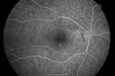
69 Year Old Woman Normal Fluorescein Angiogram786 views69-year-old woman OD 20/50 - OS 20/30 ATYPICAL PHOTOPSIAS
(0 votes)
|
|
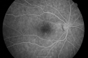
69 Year Old Woman Normal Fluorescein Angiogram790 views69-year-old woman OD 20/50 - OS 20/30 ATYPICAL PHOTOPSIAS
(0 votes)
|
|
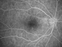
69 Year Old Woman Normal Fluorescein Angiogram753 views69-year-old woman OD 20/50 - OS 20/30 ATYPICAL PHOTOPSIAS
(0 votes)
|
|
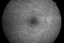
69 Year Old Woman Normal Fluorescein Angiogram747 views69-year-old woman OD 20/50 - OS 20/30 ATYPICAL PHOTOPSIAS
(0 votes)
|
|
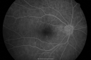
69 Year Old Woman Normal Fluorescein Angiogram677 views69-year-old woman OD 20/50 - OS 20/30 ATYPICAL PHOTOPSIAS
(0 votes)
|
|

69 Year Old Woman Normal Fundus Photograph977 views69-year-old woman OD 20/50 - OS 20/30 ATYPICAL PHOTOPSIAS
(0 votes)
|
|

69 Year Old Woman Normal Fundus Photograph867 views69-year-old woman OD 20/50 - OS 20/30 ATYPICAL PHOTOPSIAS
(0 votes)
|
|
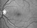
69 Year Old Woman Normal Red Free977 views69-year-old woman OD 20/50 - OS 20/30 ATYPICAL PHOTOPSIAS
(0 votes)
|
|
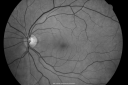
69 Year Old Woman Normal FA751 views69-year-old woman OD 20/50 - OS 20/30 ATYPICAL PHOTOPSIAS
(0 votes)
|
|
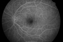
69 Year Old Woman Normal fA997 views69-year-old woman OD 20/50 - OS 20/30 ATYPICAL PHOTOPSIAS
(0 votes)
|
|

69 Year Old Woman Normal FA757 views69-year-old woman OD 20/50 - OS 20/30 ATYPICAL PHOTOPSIAS
(0 votes)
|
|
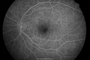
69 Year Old Woman Normal FA832 views69-year-old woman OD 20/50 - OS 20/30 ATYPICAL PHOTOPSIAS
(0 votes)
|
|
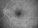
69 Year Old Woman Normal FA741 views69-year-old woman OD 20/50 - OS 20/30 ATYPICAL PHOTOPSIAS
(0 votes)
|
|
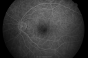
69 Year Old Woman Normal FA715 views69-year-old woman OD 20/50 - OS 20/30 ATYPICAL PHOTOPSIAS
(0 votes)
|
|
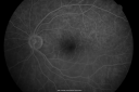
69 Year Old Woman Normal FA748 views69-year-old woman OD 20/50 - OS 20/30 ATYPICAL PHOTOPSIAS
(0 votes)
|
|
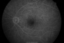
69 Year Old Woman Normal FA771 views69-year-old woman OD 20/50 - OS 20/30 ATYPICAL PHOTOPSIAS
(0 votes)
|
|
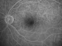
69 Year Old Woman Normal FA723 views69-year-old woman OD 20/50 - OS 20/30 ATYPICAL PHOTOPSIAS
(0 votes)
|
|
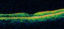
69 Year Old Woman Normal OCT898 views69-year-old woman OD 20/50 - OS 20/30 ATYPICAL PHOTOPSIAS
(0 votes)
|
|

69 Year Old Woman Normal OCT876 views69-year-old woman OD 20/50 - OS 20/30 ATYPICAL PHOTOPSIAS
(0 votes)
|
|

69 Year Old Woman Normal Fundus Photograph1130 views69-year-old woman OD 20/50 - OS 20/30 ATYPICAL PHOTOPSIAS
(0 votes)
|
|
|
|
|
|
|
69-year-old woman who I saw in the office on July 15, 2009. She had cataract surgery done in 2006, first in the left eye and subsequently in the right eye. She had vitreous separations with floaters. She also sees temporal flashing lights.
In addition she has an unusual symptom of seeing lights in both eyes, sort of blotchy lights that move around throughout the visual field that are there most of the time. They are not regionalized and they seem to be in both eyes and they are not associated with any sort of visual phenomenon, such as looking at street lights or headlights and they can happen in dim lighting or regular lighting.
VISUAL ACUITY: OD 20/50. Pinhole 20/30. OS 20/30. IOP: OD 16, OS 15. Amsler grid is normal in both eyes.
SLIT EXAMINATION: The right eye has a posterior chamber intraocular lens is in good position with clear capsular. The left eye has a posterior chamber intraocular lens is in good position with 2+ posterior capsular opacity and an open capsule center.
EXTENDED OPHTHALMOSCOPY:
OD: Vertical C/D ratio is 0.5. There is a posterior vitreous separation and trace epiretinal membrane.
OS: Vertical C/D ratio is 0.5. There is posterior vitreous separation and trace epiretinal membrane.
OCT SCAN: The OCT scan were normal as are the photos and fluorescein angiograms.
ATYPICAL PHOTOPSIAS
DISCUSSION: I explained to the patient that the photopsias she is seeing are atypical and do sound like something that we see occasionally in people who have retinal inflammatory disease, such as multiple evanescent white-dot syndrome or acute zonal occult outer retinopathy. All of those diseases though tend to have transient lights and they also tend to be regionalized. Her case is different in that she has had the lights now for several years and they seem to be diffuse.
There are some drugs which will cause retinal toxicity, such as Digitalis that can cause people to see unusual light phenomenon and that tends to be a passing thing and she is not on any medications that to my knowledge cause any retinal toxicities.
I suggested, just to be on the safe side she have a neurological evaluation, because occasionally visual symptoms are not from the eye, but from the visual pathways and it is possible that she has chronic irritation of her visual pathways somewhere.
If you have not done so, it would be reasonable to obtain a visual field, just to make sure there is not something going on with her visual pathways that might be knocking on her visual field and I told her she does not need to return here unless you or she notes further problems. Thank you for allowing me to participate in her care.