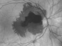
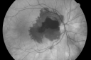
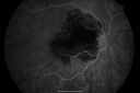
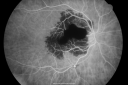
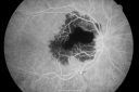
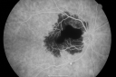
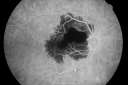
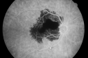
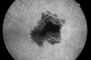
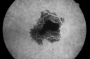
82-year-old woman was seen in the office on June 11, 2008. She had vision loss in the right eye three to four days ago. She said prior to this there was concern about a possible macular pucker in the left eye. She also had a funny unusual pain on the right side of her neck about four to five days ago, which was different than any neck pain she has had in the past. She does take Plavix. VISUAL ACUITY: OD 20/400, OS 20/40. IOP: OD 17, OS 14. SLIT LAMP EXAM: The posterior chamber intraocular lens is in good position in both eyes. EXTENDED OPHTHALMOSCOPY: OD: Vertical C/D ratio is 0.1. There is posterior vitreous separation. There is subretinal hemorrhage and a preretinal hemorrhage superior to the macula. The subretinal hemorrhage does go through the fovea. There is also a retinal tear and microaneurysm inferotemporal to the fovea. OS: Vertical C/D ratio is 0.3. There is posterior vitreous separation. There are a few patchy retinal hemorrhages inferonasal to the fovea. Photos confirm clinical findings. FLUORESCEIN ANGIOGRAM: FA shows hypofluorescence corresponding to the hemorrhage in the right eye and distinct from that there is a retinal tear and microaneurysm which is hypofluorescent inferior to the macula. The left eye has a little hyperfluorescence nasal to the fovea consistent with macular drusen. I checked her blood pressure and it was 205/94. IMPRESSION: 1. RUPTURED RETINAL ARTERIAL MACROANEURYSM – RIGHT EYE 2. HYPERTENSIVE RETINOPATHY – BOTH EYES 3. AGE-RELATED MACULAR DEGENERATION – BOTH EYES DISCUSSION: I explained to the patient that she did unfortunately have a bleed in the macula in the right eye from what looks like a ruptured retinal tear and microaneurysm. She also has a non-ruptured retinal tear and microaneurysm of the right eye as well, which hopefully will seal up on its own. I think when her blood pressure comes under better control her risk of further problems will be substantially lower. I sent her directly over to her doctors office. I asked her to return here for a check in six to eight weeks, sooner should she notice any problem. |
|||||||
|
|
|
|
||||
|
|
|
|
||||
|
|
|
|
||||
|
|
|
|
||||
|
|
|
|||||
|
|||||||
82-year-old woman was seen in the office on June 11, 2008. She had vision loss in the right eye three to four days ago. She said prior to this there was concern about a possible macular pucker in the left eye. She also had a funny unusual pain on the right side of her neck about four to five days ago, which was different than any neck pain she has had in the past. She does take Plavix. VISUAL ACUITY: OD 20/400, OS 20/40. IOP: OD 17, OS 14. SLIT LAMP EXAM: The posterior chamber intraocular lens is in good position in both eyes. EXTENDED OPHTHALMOSCOPY: OD: Vertical C/D ratio is 0.1. There is posterior vitreous separation. There is subretinal hemorrhage and a preretinal hemorrhage superior to the macula. The subretinal hemorrhage does go through the fovea. There is also a retinal tear and microaneurysm inferotemporal to the fovea. OS: Vertical C/D ratio is 0.3. There is posterior vitreous separation. There are a few patchy retinal hemorrhages inferonasal to the fovea. Photos confirm clinical findings. FLUORESCEIN ANGIOGRAM: FA shows hypofluorescence corresponding to the hemorrhage in the right eye and distinct from that there is a retinal tear and microaneurysm which is hypofluorescent inferior to the macula. The left eye has a little hyperfluorescence nasal to the fovea consistent with macular drusen. I checked her blood pressure and it was 205/94. IMPRESSION: 1. RUPTURED RETINAL ARTERIAL MACROANEURYSM – RIGHT EYE 2. HYPERTENSIVE RETINOPATHY – BOTH EYES 3. AGE-RELATED MACULAR DEGENERATION – BOTH EYES DISCUSSION: I explained to the patient that she did unfortunately have a bleed in the macula in the right eye from what looks like a ruptured retinal tear and microaneurysm. She also has a non-ruptured retinal tear and microaneurysm of the right eye as well, which hopefully will seal up on its own. I think when her blood pressure comes under better control her risk of further problems will be substantially lower. I sent her directly over to her doctors office. I asked her to return here for a check in six to eight weeks, sooner should she notice any problem.