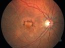
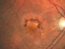
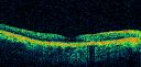
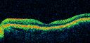
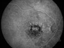
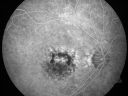
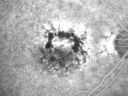
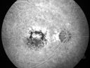
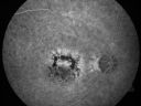
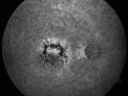
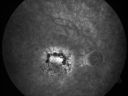
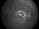
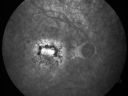
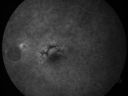
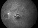
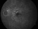
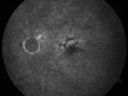
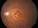
79-year-old woman was seen in the office on 12/22/2009. She has noticed, since the YAG capsulotomy in November of 2009, distortion in the right eye. Part of what she saw were fluffy blue clouds but sometimes there are gray clouds and then she notices that lines are wavy. She does not notice it as much on the Amsler grid as she does on her blinds. This is all in the right eye. The left eye continues to see fine. She does have health problems, including atrial fibrillation for which she takes Coumadin. She also notices pain around the right eye when she bends down VISUAL ACUITY: Vision OD is 20/30, OS is 20/20. IOP: OD 14, OS 13. AMSLER GRID: Right eye has central metamorphopsia. The left eye is normal. SLIT LAMP EXAM: She does have a posterior chamber intraocular lens in good position in both eyes with open capsules with 2+ vitreous debris. EXTENDED OPHTHALMOSCOPY: OD: Vertical C/D ratio is 0.4. There is no posterior vitreous separation. There are hyperpigmented spots superior to the fovea. The fovea is slightly atrophic. OS: Vertical C/D ratio is 0.4. There is no posterior vitreous separation. There are hyperpigmented spots primarily superior to the fovea. There is no intraretinal or subretinal fluid. OCT SCAN: The OCT scan confirms both macula to be dry. PHOTOGRAPHS: Photos confirm clinical findings. FLUORESCEIN ANGIOGRAPHY: There is increase in hyperfluorescence and hypofluorescence in the macula of both eyes. In the right eye there is substantially more hyperfluorescence centrally where the atrophy is. IMPRESSION: 1. NONGEOGRAPHIC PIGMENTED EPITHELIAL ATROPHY – RIGHT EYE 2. PATTERN DYSTROPHY-TYPE MACULAR DEGENERATION – BOTH EYES 3. RECENT AMSLER GRID CHANGE – RIGHT EYE 4. PAIN AROUND RIGHT EYE DISCUSSION: I explained to the patient that both maculae have pattern dystrophy-type dry age-related macular degeneration. I do not see any sign of bleeding or fluid or anything acute that needs attention. |
|||||||
|
|
|
|
||||
|
|
|
|
||||
|
|
|
|
||||
|
|
|
|
||||
|
|
|
|
||||
|
|
|
|||||
|
|||||||
79-year-old woman was seen in the office on 12/22/2009. She has noticed, since the YAG capsulotomy in November of 2009, distortion in the right eye. Part of what she saw were fluffy blue clouds but sometimes there are gray clouds and then she notices that lines are wavy. She does not notice it as much on the Amsler grid as she does on her blinds. This is all in the right eye. The left eye continues to see fine. She does have health problems, including atrial fibrillation for which she takes Coumadin. She also notices pain around the right eye when she bends down VISUAL ACUITY: Vision OD is 20/30, OS is 20/20. IOP: OD 14, OS 13. AMSLER GRID: Right eye has central metamorphopsia. The left eye is normal. SLIT LAMP EXAM: She does have a posterior chamber intraocular lens in good position in both eyes with open capsules with 2+ vitreous debris. EXTENDED OPHTHALMOSCOPY: OD: Vertical C/D ratio is 0.4. There is no posterior vitreous separation. There are hyperpigmented spots superior to the fovea. The fovea is slightly atrophic. OS: Vertical C/D ratio is 0.4. There is no posterior vitreous separation. There are hyperpigmented spots primarily superior to the fovea. There is no intraretinal or subretinal fluid. OCT SCAN: The OCT scan confirms both macula to be dry. PHOTOGRAPHS: Photos confirm clinical findings. FLUORESCEIN ANGIOGRAPHY: There is increase in hyperfluorescence and hypofluorescence in the macula of both eyes. In the right eye there is substantially more hyperfluorescence centrally where the atrophy is. IMPRESSION: 1. NONGEOGRAPHIC PIGMENTED EPITHELIAL ATROPHY – RIGHT EYE 2. PATTERN DYSTROPHY-TYPE MACULAR DEGENERATION – BOTH EYES 3. RECENT AMSLER GRID CHANGE – RIGHT EYE 4. PAIN AROUND RIGHT EYE DISCUSSION: I explained to the patient that both maculae have pattern dystrophy-type dry age-related macular degeneration. I do not see any sign of bleeding or fluid or anything acute that needs attention.