90-year-old man who I saw in the office on September 10, 2009. He recently moved to Florida from New York because his wife passed away. He had cataract surgery a little over a year ago and since the time of the cataract surgery he notices his vision in both eyes of his eyes is poor to the point where he is no longer able to read. His old chart does document that he has had branch retinal vein occlusions in both eyes in the past. It is a little difficult to sort out how long his vision has been poor. It is possible his vision in the right eye has been poor for as much as ten years. Some of the vision loss they feel previous to the cataract surgery might have been from the cataract, but it sounds like some of it might have been also from the retinal vascular occlusions.
VISUAL ACUITY: OD 20/200, OS 20/200. IOP: OD 11, OS 10.
SLIT EXAMINATION: The posterior chamber intraocular lens is in good position in both eyes. The left eye has a 2+ posterior capsular opacity.
EXTENDED OPHTHALMOSCOPY:
OD: Vertical C/D ratio is 0.2. There is an inferotemporal macular branch retinal vein occlusion. There is a well-defined, almost perfectly circular ring of pigment epithelial atrophy centrally, which is something you would see in someone who has had chronic severe cystoid macular edema for a long time.
OS: Vertical C/D ratio is 0.2. There is a pigment spot under the fovea, which is hyperpigmented possibly from macular degeneration, but also from chronic macular edema. There is a superotemporal branch retinal vein occlusion affecting an area just inside the arcades. There are a few patchy retinal hemorrhages in the periphery.
OCT SCAN: The OCT scan shows in the right eye retinal atrophy. The left eye shows macular edema. Photos confirm clinical findings.
FLUORESCEIN ANGIOGRAPHY: Fluorescein angiography confirms a branch retinal vein occlusion in both eyes with severe capillary non profusion of the inferior half of the macula in the right eye and the superior half of the macula in the left eye. There is late leakage into the fovea in the left eye.
IMPRESSION:
1. CYSTOID MACULAR EDEMA – LEFT EYE
2. MACULAR BRANCH RETINAL VEIN OCCLUSION – LEFT EYE
3. DIABETIC RETINOPATHY – LEFT EYE
4. MACULAR SCAR – RIGHT EYE
5. BRANCH RETINAL VEIN OCCLUSION – RIGHT EYE
DISCUSSION: I explained to the patient unfortunately the macula has scarring and atrophy and permanent vision loss. The left eye with the macular edema though may be responsive to treatment. I suggest that we try intravitreal Kenalog today, which I injected .05 cc. I asked him to return for a check in one month, depending on how he does it may be worthwhile considering it in the future, focal laser to that area of leakage.
|
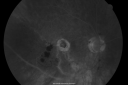
New BRVO Left Eye Old BRVO Right Eye - Right Eye Showing Bulls Eye Atrophy from Chronic CME and Non-Perfusion Infero-temporallyvu 672 fois90-year-old man decreased vision maybe one year since cataract surgery he is no longer able to read. OD 20/200, OS 20/200
(0 votes)
|
|
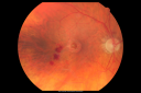
New BRVO Left Eye Old BRVO Right Eye - Right Eye Showing Bulls Eye Atrophy from Chronic CME and Non-Perfusion Infero-temporallyvu 789 fois90-year-old man decreased vision maybe one year since cataract surgery he is no longer able to read. OD 20/200, OS 20/200
(0 votes)
|
|
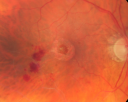
New BRVO Left Eye Old BRVO Right Eye - Right Eye Showing Bulls Eye Atrophy from Chronic CME and Non-Perfusion Infero-temporallyvu 756 fois90-year-old man decreased vision maybe one year since cataract surgery he is no longer able to read. OD 20/200, OS 20/200
(0 votes)
|
|
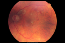
New BRVO Left Eye Old BRVO Right Eye - Left Eye Shows Twig Superior Vein Occlusion with Edemavu 730 fois90-year-old man decreased vision maybe one year since cataract surgery he is no longer able to read. OD 20/200, OS 20/200
(0 votes)
|
|

New BRVO Left Eye Old BRVO Right Eye - Left Eye Shows Twig Superior Vein Occlusion with Edemavu 787 fois90-year-old man decreased vision maybe one year since cataract surgery he is no longer able to read. OD 20/200, OS 20/200
(0 votes)
|
|
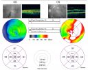
New BRVO Left Eye Old BRVO Right Eye - Left Eye Shows Twig Superior Vein Occlusion with Edemavu 571 fois90-year-old man decreased vision maybe one year since cataract surgery he is no longer able to read. OD 20/200, OS 20/200
(0 votes)
|
|

New BRVO Left Eye Old BRVO Right Eye - Right Eye Showing Bulls Eye Atrophy from Chronic CME and Non-Perfusion Infero-temporallyvu 622 fois90-year-old man decreased vision maybe one year since cataract surgery he is no longer able to read. OD 20/200, OS 20/200
(0 votes)
|
|
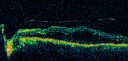
New BRVO Left Eye Old BRVO Right Eye - Left Eye Shows Twig Superior Vein Occlusion with Edemavu 566 fois90-year-old man decreased vision maybe one year since cataract surgery he is no longer able to read. OD 20/200, OS 20/200
(0 votes)
|
|
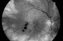
New BRVO Left Eye Old BRVO Right Eye - Right Eye Showing Bulls Eye Atrophy from Chronic CME and Non-Perfusion Infero-temporallyvu 568 fois90-year-old man decreased vision maybe one year since cataract surgery he is no longer able to read. OD 20/200, OS 20/200
(0 votes)
|
|
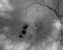
New BRVO Left Eye Old BRVO Right Eye - Right Eye Showing Bulls Eye Atrophy from Chronic CME and Non-Perfusion Infero-temporallyvu 532 fois90-year-old man decreased vision maybe one year since cataract surgery he is no longer able to read. OD 20/200, OS 20/200
(0 votes)
|
|
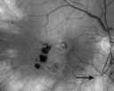
New BRVO Left Eye Old BRVO Right Eye - Right Eye Showing Bulls Eye Atrophy from Chronic CME and Non-Perfusion Infero-temporallyvu 555 fois90-year-old man decreased vision maybe one year since cataract surgery he is no longer able to read. OD 20/200, OS 20/200
(0 votes)
|
|
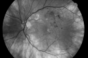
New BRVO Left Eye Old BRVO Right Eye vu 511 fois90-year-old man decreased vision maybe one year since cataract surgery he is no longer able to read. OD 20/200, OS 20/200
(0 votes)
|
|
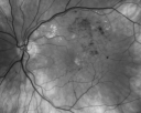
New BRVO Left Eye Old BRVO Right Eye - Left Eye Shows Twig Superior Vein Occlusion with Edemavu 532 fois90-year-old man decreased vision maybe one year since cataract surgery he is no longer able to read. OD 20/200, OS 20/200
(0 votes)
|
|

New BRVO Left Eye Old BRVO Right Eye - Left Eye Shows Twig Superior Vein Occlusion with Edemavu 645 fois90-year-old man decreased vision maybe one year since cataract surgery he is no longer able to read. OD 20/200, OS 20/200
(0 votes)
|
|
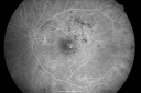
New BRVO Left Eye Old BRVO Right Eye - Left Eye Shows Twig Superior Vein Occlusion with Edemavu 598 fois90-year-old man decreased vision maybe one year since cataract surgery he is no longer able to read. OD 20/200, OS 20/200
(0 votes)
|
|
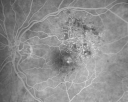
New BRVO Left Eye Old BRVO Right Eye - Left Eye Shows Twig Superior Vein Occlusion with Edemavu 488 fois90-year-old man decreased vision maybe one year since cataract surgery he is no longer able to read. OD 20/200, OS 20/200
(0 votes)
|
|
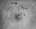
New BRVO Left Eye Old BRVO Right Eye - Left Eye Shows Twig Superior Vein Occlusion with Edema Arrow Shows Site of Occlusionvu 603 fois90-year-old man decreased vision maybe one year since cataract surgery he is no longer able to read. OD 20/200, OS 20/200
(0 votes)
|
|
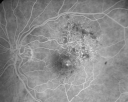
New BRVO Left Eye Old BRVO Right Eye - Left Eye Shows Twig Superior Vein Occlusion with Edemavu 513 fois90-year-old man decreased vision maybe one year since cataract surgery he is no longer able to read. OD 20/200, OS 20/200
(0 votes)
|
|
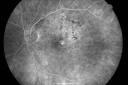
New BRVO Left Eye Old BRVO Right Eye - Left Eye Shows Twig Superior Vein Occlusion with Edemavu 498 fois90-year-old man decreased vision maybe one year since cataract surgery he is no longer able to read. OD 20/200, OS 20/200
(0 votes)
|
|
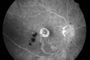
New BRVO Left Eye Old BRVO Right Eye - Right Eye Showing Bulls Eye Atrophy from Chronic CME and Non-Perfusion Infero-temporallyvu 632 fois90-year-old man decreased vision maybe one year since cataract surgery he is no longer able to read. OD 20/200, OS 20/200
(0 votes)
|
|
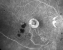
New BRVO Left Eye Old BRVO Right Eye - Right Eye Showing Bulls Eye Atrophy from Chronic CME and Non-Perfusion Infero-temporallyvu 495 fois90-year-old man decreased vision maybe one year since cataract surgery he is no longer able to read. OD 20/200, OS 20/200
(0 votes)
|
|
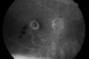
New BRVO Left Eye Old BRVO Right Eye - Right Eye Showing Bulls Eye Atrophy from Chronic CME and Non-Perfusion Infero-temporallyvu 581 fois90-year-old man decreased vision maybe one year since cataract surgery he is no longer able to read. OD 20/200, OS 20/200
(0 votes)
|
|

New BRVO Left Eye Old BRVO Right Eye - Left Eye Shows Twig Superior Vein Occlusion with Edemavu 578 fois90-year-old man decreased vision maybe one year since cataract surgery he is no longer able to read. OD 20/200, OS 20/200
(0 votes)
|
|
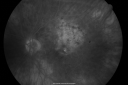
New BRVO Left Eye Old BRVO Right Eye - Left Eye Shows Twig Superior Vein Occlusion with Edemavu 564 fois90-year-old man decreased vision maybe one year since cataract surgery he is no longer able to read. OD 20/200, OS 20/200
(0 votes)
|
|
|
|
90-year-old man who I saw in the office on September 10, 2009. He recently moved to Florida from New York because his wife passed away. He had cataract surgery a little over a year ago and since the time of the cataract surgery he notices his vision in both eyes of his eyes is poor to the point where he is no longer able to read. His old chart does document that he has had branch retinal vein occlusions in both eyes in the past. It is a little difficult to sort out how long his vision has been poor. It is possible his vision in the right eye has been poor for as much as ten years. Some of the vision loss they feel previous to the cataract surgery might have been from the cataract, but it sounds like some of it might have been also from the retinal vascular occlusions.
VISUAL ACUITY: OD 20/200, OS 20/200. IOP: OD 11, OS 10.
SLIT EXAMINATION: The posterior chamber intraocular lens is in good position in both eyes. The left eye has a 2+ posterior capsular opacity.
EXTENDED OPHTHALMOSCOPY:
OD: Vertical C/D ratio is 0.2. There is an inferotemporal macular branch retinal vein occlusion. There is a well-defined, almost perfectly circular ring of pigment epithelial atrophy centrally, which is something you would see in someone who has had chronic severe cystoid macular edema for a long time.
OS: Vertical C/D ratio is 0.2. There is a pigment spot under the fovea, which is hyperpigmented possibly from macular degeneration, but also from chronic macular edema. There is a superotemporal branch retinal vein occlusion affecting an area just inside the arcades. There are a few patchy retinal hemorrhages in the periphery.
OCT SCAN: The OCT scan shows in the right eye retinal atrophy. The left eye shows macular edema. Photos confirm clinical findings.
FLUORESCEIN ANGIOGRAPHY: Fluorescein angiography confirms a branch retinal vein occlusion in both eyes with severe capillary non profusion of the inferior half of the macula in the right eye and the superior half of the macula in the left eye. There is late leakage into the fovea in the left eye.
IMPRESSION:
1. CYSTOID MACULAR EDEMA – LEFT EYE
2. MACULAR BRANCH RETINAL VEIN OCCLUSION – LEFT EYE
3. DIABETIC RETINOPATHY – LEFT EYE
4. MACULAR SCAR – RIGHT EYE
5. BRANCH RETINAL VEIN OCCLUSION – RIGHT EYE
DISCUSSION: I explained to the patient unfortunately the macula has scarring and atrophy and permanent vision loss. The left eye with the macular edema though may be responsive to treatment. I suggest that we try intravitreal Kenalog today, which I injected .05 cc. I asked him to return for a check in one month, depending on how he does it may be worthwhile considering it in the future, focal laser to that area of leakage.


