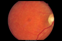
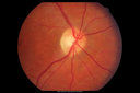
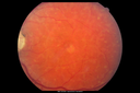
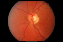
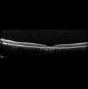
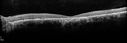
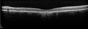
Stargardt's Macular Dystrophy - Heterozygous ABCA4 T>C substitution at potisoin -10 of intro 38
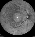
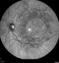
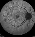
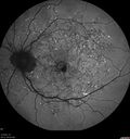
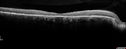
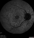
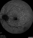
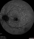
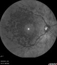
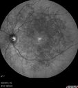
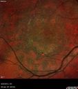
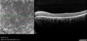
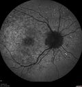
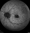
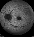
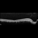
65 year old woman with gradually declining central vision. VA 20/200 in both eyes in the later photos (left eye was better in earlier photos). FAF shows pisciform tri-radiate lesions that are hyper FAF typical of stargardt's fundus flavimaculatus
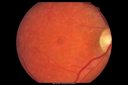
Stargardt's Macular Dystrophy ABCA4 positivevaatamisi: 594Images show some progression over 3 years of macular dystrophy. (0 hinnangut)
Stargardt's Macular Dystrophy ABCA4 positivevaatamisi: 536Images show some progression over 3 years of macular dystrophy. (0 hinnangut)
Stargardt's Macular Dystrophy ABCA4 positivevaatamisi: 634Images show some progression over 3 years of macular dystrophy. (0 hinnangut)
Stargardt's Macular Dystrophy ABCA4 positivevaatamisi: 604Images show some progression over 3 years of macular dystrophy. (0 hinnangut)
Stargardt's Macular Dystrophy ABCA4 positivevaatamisi: 553Images show some progression over 3 years of macular dystrophy. (0 hinnangut)
Stargardt's Macular Dystrophy ABCA4 positivevaatamisi: 655Images show some progression over 3 years of macular dystrophy. (0 hinnangut)
Stargardt's Macular Dystrophy ABCA4 positivevaatamisi: 551Images show some progression over 3 years of macular dystrophy. (0 hinnangut)
Stargardt's Macular Dystrophy ABCA4 positivevaatamisi: 597 Fundus Autofluorescence shows central atrophy with Hyper FAF pisciform triradiate lesions. Images show some progression over 3 years of macular dystrophy. (0 hinnangut)
Stargardt's Macular Dystrophy ABCA4 positivevaatamisi: 566 Fundus Autofluorescence shows central atrophy with Hyper FAF pisciform triradiate lesions. Images show some progression over 3 years of macular dystrophy. (0 hinnangut)
Stargardt's Macular Dystrophy ABCA4 positivevaatamisi: 480 Fundus Autofluorescence shows central atrophy with Hyper FAF pisciform triradiate lesions. Images show some progression over 3 years of macular dystrophy. (0 hinnangut)
Stargardt's Macular Dystrophy ABCA4 positivevaatamisi: 452Images show some progression over 3 years of macular dystrophy. (0 hinnangut)
Stargardt's Macular Dystrophy ABCA4 positivevaatamisi: 490Images show some progression over 3 years of macular dystrophy. (0 hinnangut)
Stargardt's Macular Dystrophy ABCA4 positivevaatamisi: 585 Fundus Autofluorescence shows central atrophy with Hyper FAF pisciform triradiate lesions. Images show some progression over 3 years of macular dystrophy. (0 hinnangut)
Stargardt's Macular Dystrophy ABCA4 positivevaatamisi: 519 Fundus Autofluorescence shows central atrophy with Hyper FAF pisciform triradiate lesions. Images show some progression over 3 years of macular dystrophy. (0 hinnangut)
Stargardt's Macular Dystrophy ABCA4 positivevaatamisi: 496 Fundus Autofluorescence shows central atrophy with Hyper FAF pisciform triradiate lesions. Images show some progression over 3 years of macular dystrophy. (0 hinnangut)
Stargardt's Macular Dystrophy ABCA4 positivevaatamisi: 450Images show some progression over 3 years of macular dystrophy. (0 hinnangut)
Stargardt's Macular Dystrophy ABCA4 positivevaatamisi: 490Images show some progression over 3 years of macular dystrophy. (0 hinnangut)
Stargardt's Macular Dystrophy ABCA4 positivevaatamisi: 442Images show some progression over 3 years of macular dystrophy. (0 hinnangut)
Stargardt's Macular Dystrophy ABCA4 positivevaatamisi: 508Images show some progression over 3 years of macular dystrophy. (0 hinnangut)
Stargardt's Macular Dystrophy ABCA4 positivevaatamisi: 499Images show some progression over 3 years of macular dystrophy. (0 hinnangut)
Stargardt's Macular Dystrophy ABCA4 positivevaatamisi: 494Images show some progression over 3 years of macular dystrophy. (0 hinnangut)
Stargardt's Macular Dystrophy ABCA4 positivevaatamisi: 502 Fundus Autofluorescence shows central atrophy with Hyper FAF pisciform triradiate lesions. Images show some progression over 3 years of macular dystrophy. (0 hinnangut)
Stargardt's Macular Dystrophy ABCA4 positivevaatamisi: 530 Fundus Autofluorescence shows central atrophy with Hyper FAF pisciform triradiate lesions. Images show some progression over 3 years of macular dystrophy. (0 hinnangut)
Stargardt's Macular Dystrophy ABCA4 positivevaatamisi: 538 Fundus Autofluorescence shows central atrophy with Hyper FAF pisciform triradiate lesions. Images show some progression over 3 years of macular dystrophy. (0 hinnangut)
Stargardt's Macular Dystrophy ABCA4 positivevaatamisi: 503Images show some progression over 3 years of macular dystrophy. (0 hinnangut)
Stargardt's Macular Dystrophy ABCA4 positivevaatamisi: 500Images show some progression over 3 years of macular dystrophy. (0 hinnangut)
Stargardt's Macular Dystrophy ABCA4 positivevaatamisi: 465Images show some progression over 3 years of macular dystrophy. (0 hinnangut)
Stargardt's Macular Dystrophy ABCA4 positivevaatamisi: 501Images show some progression over 3 years of macular dystrophy. (0 hinnangut)
28 faili 1 lehel
65 year old woman with gradually declining central vision. VA 20/200 in both eyes in the later photos (left eye was better in earlier photos). FAF shows pisciform tri-radiate lesions that are hyper FAF typical of stargardt's fundus flavimaculatus