Acute Branch Retinal Artery Occlusion (fellow eye has old BRVO)
|
|
84 year old woman. Two days ago she noticed a shadow in her inferior vision. This was not associated with any focal neurological changes. She denies any headaches.
•VA OD: cc20/50-1 PHNI
•VA OS: cc20/50+2 PH20/40-2 The left eye has a white area visible on photography, the OCT shows inner retinal edema and the FA shows poor circulation.
|
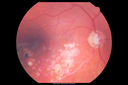
Acute Branch Retinal Artery Occlusion Left Eyevista 759 vecesThis 84 year old woman had an old BRVO in the right eye and saw a shadow for 2 days in the left eye. She has a superotemporal BRAO. The photos show retinal whitening, the OCT shows inner retinal edema and the FA shows poor circulation.
(0 votos)
|
|

Acute Branch Retinal Artery Occlusion Left Eyevista 631 vecesThis 84 year old woman had an old BRVO in the right eye and saw a shadow for 2 days in the left eye. She has a superotemporal BRAO. The photos show retinal whitening, the OCT shows inner retinal edema and the FA shows poor circulation.
(0 votos)
|
|

Acute Branch Retinal Artery Occlusion Left Eyevista 702 vecesThis 84 year old woman had an old BRVO in the right eye and saw a shadow for 2 days in the left eye. She has a superotemporal BRAO. The photos show retinal whitening, the OCT shows inner retinal edema and the FA shows poor circulation.
(0 votos)
|
|

Acute Branch Retinal Artery Occlusion Left Eyevista 690 vecesThis 84 year old woman had an old BRVO in the right eye and saw a shadow for 2 days in the left eye. She has a superotemporal BRAO. The photos show retinal whitening, the OCT shows inner retinal edema and the FA shows poor circulation.
(0 votos)
|
|

Acute Branch Retinal Artery Occlusion Left Eyevista 677 vecesThis 84 year old woman had an old BRVO in the right eye and saw a shadow for 2 days in the left eye. She has a superotemporal BRAO. The photos show retinal whitening, the OCT shows inner retinal edema and the FA shows poor circulation.
(0 votos)
|
|

Acute Branch Retinal Artery Occlusion Left Eyevista 667 vecesThis 84 year old woman had an old BRVO in the right eye and saw a shadow for 2 days in the left eye. She has a superotemporal BRAO. The photos show retinal whitening, the OCT shows inner retinal edema and the FA shows poor circulation.
(0 votos)
|
|
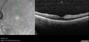
Acute Branch Retinal Artery Occlusion Left Eyevista 495 vecesThis 84 year old woman had an old BRVO in the right eye and saw a shadow for 2 days in the left eye. She has a superotemporal BRAO. The photos show retinal whitening, the OCT shows inner retinal edema and the FA shows poor circulation.
(0 votos)
|
|

Acute Branch Retinal Artery Occlusion Left Eyevista 514 vecesThis 84 year old woman had an old BRVO in the right eye and saw a shadow for 2 days in the left eye. She has a superotemporal BRAO. The photos show retinal whitening, the OCT shows inner retinal edema and the FA shows poor circulation.
(0 votos)
|
|

Acute Branch Retinal Artery Occlusion Left Eyevista 518 vecesThis 84 year old woman had an old BRVO in the right eye and saw a shadow for 2 days in the left eye. She has a superotemporal BRAO. The photos show retinal whitening, the OCT shows inner retinal edema and the FA shows poor circulation.
(0 votos)
|
|

Acute Branch Retinal Artery Occlusion Left Eyevista 592 vecesThis 84 year old woman had an old BRVO in the right eye and saw a shadow for 2 days in the left eye. She has a superotemporal BRAO. The photos show retinal whitening, the OCT shows inner retinal edema and the FA shows poor circulation.
(0 votos)
|
|

Acute Branch Retinal Artery Occlusion Left Eyevista 508 vecesThis 84 year old woman had an old BRVO in the right eye and saw a shadow for 2 days in the left eye. She has a superotemporal BRAO. The photos show retinal whitening, the OCT shows inner retinal edema and the FA shows poor circulation.
(0 votos)
|
|

Acute Branch Retinal Artery Occlusion Left Eyevista 452 vecesThis 84 year old woman had an old BRVO in the right eye and saw a shadow for 2 days in the left eye. She has a superotemporal BRAO. The photos show retinal whitening, the OCT shows inner retinal edema and the FA shows poor circulation.
(0 votos)
|
|

Acute Branch Retinal Artery Occlusion Left Eyevista 523 vecesThis 84 year old woman had an old BRVO in the right eye and saw a shadow for 2 days in the left eye. She has a superotemporal BRAO. The photos show retinal whitening, the OCT shows inner retinal edema and the FA shows poor circulation.
(0 votos)
|
|

Acute Branch Retinal Artery Occlusion Left Eyevista 593 vecesThis 84 year old woman had an old BRVO in the right eye and saw a shadow for 2 days in the left eye. She has a superotemporal BRAO. The photos show retinal whitening, the OCT shows inner retinal edema and the FA shows poor circulation.
(0 votos)
|
|

Acute Branch Retinal Artery Occlusion Left Eyevista 638 vecesThis 84 year old woman had an old BRVO in the right eye and saw a shadow for 2 days in the left eye. She has a superotemporal BRAO. The photos show retinal whitening, the OCT shows inner retinal edema and the FA shows poor circulation.
(0 votos)
|
|

Acute Branch Retinal Artery Occlusion Left Eyevista 672 vecesThis 84 year old woman had an old BRVO in the right eye and saw a shadow for 2 days in the left eye. She has a superotemporal BRAO. The photos show retinal whitening, the OCT shows inner retinal edema and the FA shows poor circulation.
(0 votos)
|
|

Acute Branch Retinal Artery Occlusion Left Eyevista 652 vecesThis 84 year old woman had an old BRVO in the right eye and saw a shadow for 2 days in the left eye. She has a superotemporal BRAO. The photos show retinal whitening, the OCT shows inner retinal edema and the FA shows poor circulation.
(0 votos)
|
|

Acute Branch Retinal Artery Occlusion Left Eyevista 687 vecesThis 84 year old woman had an old BRVO in the right eye and saw a shadow for 2 days in the left eye. She has a superotemporal BRAO. The photos show retinal whitening, the OCT shows inner retinal edema and the FA shows poor circulation.
(0 votos)
|
|

Acute Branch Retinal Artery Occlusion Left Eyevista 561 vecesThis 84 year old woman had an old BRVO in the right eye and saw a shadow for 2 days in the left eye. She has a superotemporal BRAO. The photos show retinal whitening, the OCT shows inner retinal edema and the FA shows poor circulation.
(0 votos)
|
|

Acute Branch Retinal Artery Occlusion Left Eyevista 682 vecesThis 84 year old woman had an old BRVO in the right eye and saw a shadow for 2 days in the left eye. She has a superotemporal BRAO. The photos show retinal whitening, the OCT shows inner retinal edema and the FA shows poor circulation.
(0 votos)
|
|

Acute Branch Retinal Artery Occlusion Left Eyevista 571 vecesThis 84 year old woman had an old BRVO in the right eye and saw a shadow for 2 days in the left eye. She has a superotemporal BRAO. The photos show retinal whitening, the OCT shows inner retinal edema and the FA shows poor circulation.
(0 votos)
|
|

Acute Branch Retinal Artery Occlusion Left Eyevista 526 vecesThis 84 year old woman had an old BRVO in the right eye and saw a shadow for 2 days in the left eye. She has a superotemporal BRAO. The photos show retinal whitening, the OCT shows inner retinal edema and the FA shows poor circulation.
(0 votos)
|
|

Acute Branch Retinal Artery Occlusion Left Eyevista 550 vecesThis 84 year old woman had an old BRVO in the right eye and saw a shadow for 2 days in the left eye. She has a superotemporal BRAO. The photos show retinal whitening, the OCT shows inner retinal edema and the FA shows poor circulation.
(0 votos)
|
|
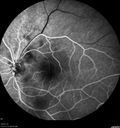
Acute Branch Retinal Artery Occlusion Left Eyevista 550 vecesThis 84 year old woman had an old BRVO in the right eye and saw a shadow for 2 days in the left eye. She has a superotemporal BRAO. The photos show retinal whitening, the OCT shows inner retinal edema and the FA shows poor circulation.
(0 votos)
|
|

Acute Branch Retinal Artery Occlusion Left Eyevista 567 vecesThis 84 year old woman had an old BRVO in the right eye and saw a shadow for 2 days in the left eye. She has a superotemporal BRAO. The photos show retinal whitening, the OCT shows inner retinal edema and the FA shows poor circulation.
(0 votos)
|
|

Acute Branch Retinal Artery Occlusion Left Eyevista 578 vecesThis 84 year old woman had an old BRVO in the right eye and saw a shadow for 2 days in the left eye. She has a superotemporal BRAO. The photos show retinal whitening, the OCT shows inner retinal edema and the FA shows poor circulation.
(0 votos)
|
|

Acute Branch Retinal Artery Occlusion Left Eyevista 557 vecesThis 84 year old woman had an old BRVO in the right eye and saw a shadow for 2 days in the left eye. She has a superotemporal BRAO. The photos show retinal whitening, the OCT shows inner retinal edema and the FA shows poor circulation.
(0 votos)
|
|

Acute Branch Retinal Artery Occlusion Left Eyevista 569 vecesThis 84 year old woman had an old BRVO in the right eye and saw a shadow for 2 days in the left eye. She has a superotemporal BRAO. The photos show retinal whitening, the OCT shows inner retinal edema and the FA shows poor circulation.
(0 votos)
|
|
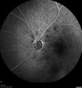
Acute Branch Retinal Artery Occlusion Left Eyevista 673 vecesThis 84 year old woman had an old BRVO in the right eye and saw a shadow for 2 days in the left eye. She has a superotemporal BRAO. The photos show retinal whitening, the OCT shows inner retinal edema and the FA shows poor circulation.
(0 votos)
|
|
|
|
|
| 29 archivos en 1 página(s) |
|
84 year old woman. Two days ago she noticed a shadow in her inferior vision. This was not associated with any focal neurological changes. She denies any headaches.
•VA OD: cc20/50-1 PHNI
•VA OS: cc20/50+2 PH20/40-2 The left eye has a white area visible on photography, the OCT shows inner retinal edema and the FA shows poor circulation.


